Anatomy of an Offering: CT Imaging and Kemet’s Votive Animals in British Collections
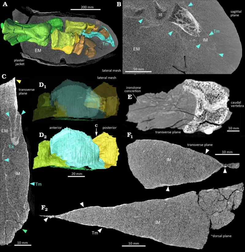
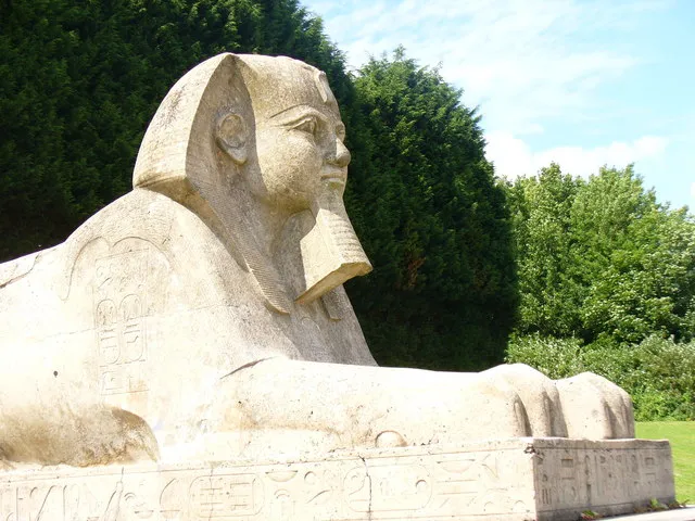
Photo: E. T. Saitta et al., CC BY 4.0, via Wikimedia Commons
On a grey November morning in 2026, the language of a hospital imaging department and the language of a museum storeroom meet over a small bundle of linen. A mummified ibis, perhaps no larger than a loaf of bread, is placed on a scanner bed. No bandage is lifted. No beak is exposed. Instead, a clinical CT system records thousands of cross-sectional images, and a conservation team waits for the anatomy of an ancient offering to appear on screen.
This is not an isolated curiosity. Across the UK, museum conservation departments have been building working relationships with clinical imaging specialists, drawing on equipment and expertise designed for human medicine to study animal mummies non-invasively. The practice suits both parties. Hospitals have sophisticated scanners, protocols and radiographers; museums have objects whose internal structures have long been guessed at but rarely seen. The result is a growing archive of digital evidence about Kemet’s votive animals, produced without the irreversible intervention that unwrapping once implied.
A clinical gaze on sacred remains
Animal mummies are among the most abundant Egyptian artefacts in British collections. Ibises, cats, dogs, falcons, shrews and other creatures were dedicated in vast numbers at temples and shrines, often as offerings on behalf of a petitioner. Victorian and Edwardian collectors brought many to Britain, where they entered national and provincial museums as curiosities, teaching specimens or archaeological material. For much of the twentieth century, study often meant unwrapping or dissection. The object became a specimen, and the wrapping a barrier to be removed.
CT imaging changes that sequence. The scanner produces a three-dimensional volume that can be rotated, sliced and measured. Radiographers can distinguish bone from textile, resin from desiccated tissue, and sometimes identify the species, the position of the body and the method of preparation. A cat may be curled tightly; an ibis may be folded with its beak along its body; a bundle may contain more than one animal, or very little animal at all. These details matter because they are evidence of craft, ritual and economy, not merely of natural history.
What the scans reveal
One of the most important contributions of CT work is the ability to separate preparation from presentation. The wrappings are carefully arranged, sometimes with painted faces or decorative bands, but the interior can tell a different story. Scans have shown that some votive bundles contain incomplete animals, mixtures of species or materials intended to give bulk and shape. Such findings should not be read simply as deception. They point to a production system in which demand was high, supply was organised, and temple workshops responded to pilgrims with varying budgets.
The scans also clarify how animals were killed, preserved and folded. Evidence of trauma, desiccation and resin treatment can be compared across collections, allowing researchers to ask whether particular temples or workshops favoured particular methods. For Kemet’s temple economies, this is valuable. Votive animals were not incidental piety; they were part of a complex system of donation, priestly income, craft specialisation and regional pilgrimage. A cat mummy from a provincial shrine and an ibis from a major cult centre may look similar under linen, but their internal anatomy and preparation can reveal different supply chains and ritual expectations.
Pilgrimage, production and the limits of inference
The temptation with any new imaging technique is to over-read. A scan is not an excavation, and a digital slice is not a social history. Researchers must be careful about sample size, provenance and the bias of collections formed by nineteenth-century acquisition. Many British-held animal mummies lack secure archaeological context. A beautifully rendered image can seem more authoritative than the fragmentary records behind it.
Even so, the cumulative evidence is useful. When CT data are combined with textual sources, temple architecture and known pilgrimage routes, patterns begin to emerge. The sheer scale of animal dedication implies organised breeding and procurement. The uniformity of certain wrappings suggests workshop conventions. The variation in internal contents suggests a market that accommodated different levels of expense. These are modest conclusions, but they are more grounded than the older assumption that animal mummies were simply mass-produced frauds or mere religious curiosities.
The ethics of non-invasive study
Non-invasive imaging is often presented as an ethical solution, and in important respects it is. It avoids destructive sampling, preserves the wrappings and allows repeated study without further handling. For collections that hold human and animal remains, that matters. British museums have become more cautious about display and intervention, and CT scanning offers a way to generate research value while reducing physical intrusion.
Yet non-invasive does not mean neutral. The decision to scan is still a decision to collect data, interpret remains and publish images. It raises questions about consent, provenance and the colonial routes by which many objects entered UK collections. Animal mummies may not carry the same legal and moral frameworks as human remains, but they are not mere things. They are ritual objects, once offered in sacred settings, and their treatment in British laboratories should be transparent. Museums should record why an object is scanned, who has access to the data and how the results will be displayed. Where possible, source communities and Egyptian heritage professionals should be part of those conversations.
A British network of expertise
The practical strength of this work lies in collaboration. Clinical scanners are expensive, and museum budgets are not. Partnerships with NHS imaging departments, university radiography programmes and specialist veterinary imaging centres allow smaller regional museums to access equipment that would otherwise be out of reach. The Ashmolean, Manchester Museum, the British Museum and numerous provincial collections have all explored such approaches, and the results are gradually feeding into conservation records and public interpretation.
For visitors, the appeal is immediate. A scan can show a wrapped object as a creature, an offering and a product of skilled labour all at once. For conservators, it is a tool for condition assessment. For historians, it is a fragment of evidence about how Kemet organised devotion. The scanner does not resolve the ethical questions surrounding these collections, but it can make them more precise. That is a modest and worthwhile advance: not unwrapping the past, but learning to see it more carefully.